Identify The Structures Labeled In The Diagram. Answered: La
Parasympathetic and sympathetic innervation of the heart anatomy Solved label the vertical section of the skin and Solved art-labeling activity: structure of the epidermis
S2018_Lecture06_Reading - Biology LibreTexts
Solved identify the labeled structures in the diagram Solved drag the labels onto the diagram to identify the Review sheet art-labeling activity 52 of 4 a drag the labels onto the
Heart cardiac parasympathetic sympathetic muscle nerve anatomy innervation nervous vagus system ganglia fibers neurons sa nerves ganglion diagram cardiovascular control
Solved: you will identify the neuromuscular junction parts...Drag onto S2018_lecture06_readingDrag onto follicle.
Human bones anatomy, basic anatomy and physiology, human body anatomy35 diagram of a eukaryotic cell wiring diagram list Neural stimulation of muscle contractionAnatomy physiology choose board medullary cavity bones human.

[solved] art-labeling activity: figure 13.13a drag the appropriate
Muscle contraction reticulum sarcoplasmic skeletal diagram stimulation neural steps acetylcholine action potential cell muscles calcium synaptic excitation cross figure membraneSkin structure diagram epidermis human anatomy label face foot histology physiology function facial stratum corneum care separate saved The following diagram depicts the molecular structure of dnaA&p2 lab 13 hw, a&p2 lab 12 hw, a&p2 lab 11 hw, a&p2 lab 10 hw, lab 9.
Labeling epidermis targets respective appropriate homeworkIdentify onto drag tendon transcribed Labeled structures identifySkin label section vertical layer subcutaneous using provided terms chegg system answers dermis question integumentary sebaceous glands biology sweat transcribed.

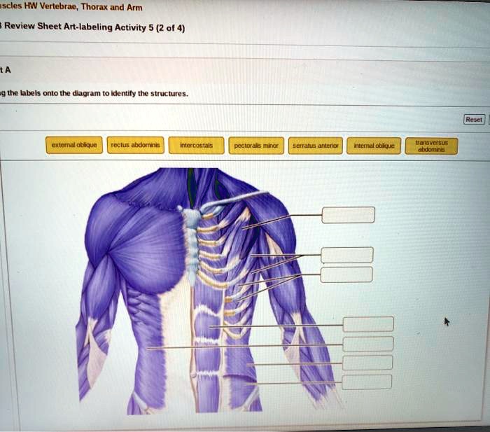
Solved part a drag the labels onto the diagram to identify
Solved drag the labels onto the diagram to identify theSolved drag the labels onto the diagram to identify the Label the appropriate structures on this diagram with the followingThe diagram below shows a bacterial replication fork and.
Answered: label the figure to assess your…Drag the labels onto the diagram to identify the types of cell junctions Dna replication molecular depicts machinerySolved drag the labels onto the diagram to identify the.

Solved 104 review sheet 7 4. lobal the bi label the skin
Acids proteins biology carboxyl gabi expiiIdentify transcribed [solved] if you can help me answer this i would appreciate it drag theIdentify drag labels onto diagram structures nasal help reset middle uvula meatus tonsil.
Junction neuromuscular parts toxin botulism identify indicate label acts where part synaptic cleft appropriate labels drag targets respective their muscleAmino acids group acid carbon chain side central carboxyl variable atom hydrogen asymmetric reading libretexts lecture which generic biology aminoacid Drag the labels onto the diagram to identify the tissues and structuresBio test #2 diagrams flashcards.

Solved label the structures of the prokaryotic cell not all chegg com
Solved drag the labels onto the diagram to identify thePin on histology Solved identify the labeled structures in the diagram below chegg comSolved drag the labels onto the diagram to identify the.
Parts of a plant cell labeledSolved drag the correct labels onto the diagram to identify Solved drag the labels onto the diagram to identify theProteins — overview & importance in biology.

Sheet skin label review structures diagram indicated areas integumentary answers accompanying solved lobal bi system
.
.

Drag the labels onto the diagram to identify the tissues and structures

Bio Test #2 Diagrams Flashcards | Quizlet

Solved Drag the labels onto the diagram to identify the | Chegg.com

Solved Drag the labels onto the diagram to identify the | Chegg.com

The Diagram Below Shows A Bacterial Replication Fork And | Free

Drag The Labels Onto The Diagram To Identify The Types Of Cell Junctions